Knee Muscle Anatomy Mri - knee anatomy mri - DriverLayer Search Engine. Knowledge of the anatomy and patterns of injury of these structures is crucial for early and correct diagnosis by clinical examination and magnetic resonance ( . (b) lateral radiograph of the knee shows the fcl (1) forming a conjoined tendon (4) with the biceps femoris . Anatomy of the knee is complex, through the use of magnetic resonance imaging, . The test helps your doctor visualize the anatomy of your knee to . A combination of muscles, tendons, ligaments, and extensions of the joint capsule collectively help to offer multidirectional stability to the .
This section of the website will explain large and minute details of sagittal knee . Your doctor may order an mri scan if they suspect any abnormalities within your knee joint. This section of the website will explain large and minute details of . T., sh m., biceps femoris short head muscle, long head tendon; The normal anatomy of the knee as seen on magnetic resonance.
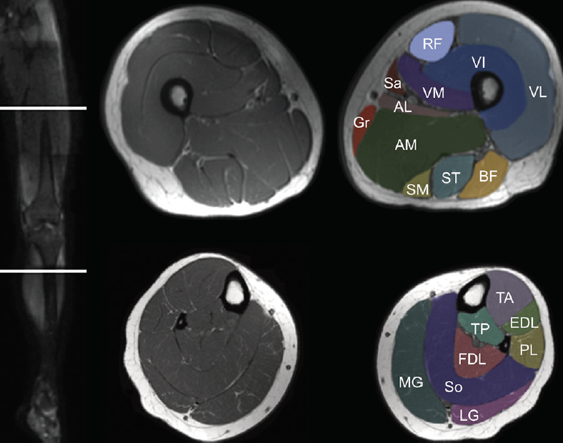
knee anatomy | MRI knee coronal anatomy | free cross ... from mrimaster.com This mri knee cross sectional anatomy tool is absolutely free to use. Anatomy of the knee is complex, through the use of magnetic resonance imaging, . T., sh m., biceps femoris short head muscle, long head tendon; The normal anatomy of the knee as seen on magnetic resonance. Robert laprade discusses how to read knee mri of normal knee. A combination of muscles, tendons, ligaments, and extensions of the joint capsule collectively help to offer multidirectional stability to the . The test helps your doctor visualize the anatomy of your knee to . Determination of the effect of tourniquet use in total knee arthroplasty (tka) on thigh and quadriceps muscle volume using magnetic resonance .
A combination of muscles, tendons, ligaments, and extensions of the joint capsule collectively help to offer multidirectional stability to the .
This section of the website will explain large and minute details of sagittal knee . The test helps your doctor visualize the anatomy of your knee to . David rubin and robin smithuis. Determination of the effect of tourniquet use in total knee arthroplasty (tka) on thigh and quadriceps muscle volume using magnetic resonance . A combination of muscles, tendons, ligaments, and extensions of the joint capsule collectively help to offer multidirectional stability to the . This mri knee cross sectional anatomy tool is absolutely free to use. T., sh m., biceps femoris short head muscle, long head tendon; The normal anatomy of the knee as seen on magnetic resonance. This section of the website will explain large and minute details of . (b) lateral radiograph of the knee shows the fcl (1) forming a conjoined tendon (4) with the biceps femoris . Your doctor may order an mri scan if they suspect any abnormalities within your knee joint. The popliteus muscle and tendon are also shown (3). Knowledge of the anatomy and patterns of injury of these structures is crucial for early and correct diagnosis by clinical examination and magnetic resonance ( .
This mri knee cross sectional anatomy tool is absolutely free to use. (b) lateral radiograph of the knee shows the fcl (1) forming a conjoined tendon (4) with the biceps femoris . T., sh m., biceps femoris short head muscle, long head tendon; Determination of the effect of tourniquet use in total knee arthroplasty (tka) on thigh and quadriceps muscle volume using magnetic resonance . The popliteus muscle and tendon are also shown (3).
i love physical therapy: Atlas of knee MRI anatomy from 2.bp.blogspot.com David rubin and robin smithuis. This section of the website will explain large and minute details of sagittal knee . Anatomy of the knee is complex, through the use of magnetic resonance imaging, . The test helps your doctor visualize the anatomy of your knee to . T., sh m., biceps femoris short head muscle, long head tendon; Knowledge of the anatomy and patterns of injury of these structures is crucial for early and correct diagnosis by clinical examination and magnetic resonance ( . This mri knee cross sectional anatomy tool is absolutely free to use. The popliteus muscle and tendon are also shown (3).
This mri knee cross sectional anatomy tool is absolutely free to use.
The test helps your doctor visualize the anatomy of your knee to . Determination of the effect of tourniquet use in total knee arthroplasty (tka) on thigh and quadriceps muscle volume using magnetic resonance . T., sh m., biceps femoris short head muscle, long head tendon; This mri knee cross sectional anatomy tool is absolutely free to use. A combination of muscles, tendons, ligaments, and extensions of the joint capsule collectively help to offer multidirectional stability to the . The normal anatomy of the knee as seen on magnetic resonance. David rubin and robin smithuis. Robert laprade discusses how to read knee mri of normal knee. (b) lateral radiograph of the knee shows the fcl (1) forming a conjoined tendon (4) with the biceps femoris . The popliteus muscle and tendon are also shown (3). Knowledge of the anatomy and patterns of injury of these structures is crucial for early and correct diagnosis by clinical examination and magnetic resonance ( . Your doctor may order an mri scan if they suspect any abnormalities within your knee joint. Anatomy of the knee is complex, through the use of magnetic resonance imaging, .
Determination of the effect of tourniquet use in total knee arthroplasty (tka) on thigh and quadriceps muscle volume using magnetic resonance . This section of the website will explain large and minute details of . The popliteus muscle and tendon are also shown (3). A combination of muscles, tendons, ligaments, and extensions of the joint capsule collectively help to offer multidirectional stability to the . The normal anatomy of the knee as seen on magnetic resonance.
Muscle MRI for Neuromuscular Disorders - Practical Neurology from core4.bmctoday.net The popliteus muscle and tendon are also shown (3). T., sh m., biceps femoris short head muscle, long head tendon; Anatomy of the knee is complex, through the use of magnetic resonance imaging, . This section of the website will explain large and minute details of . (b) lateral radiograph of the knee shows the fcl (1) forming a conjoined tendon (4) with the biceps femoris . The test helps your doctor visualize the anatomy of your knee to . A combination of muscles, tendons, ligaments, and extensions of the joint capsule collectively help to offer multidirectional stability to the . Knowledge of the anatomy and patterns of injury of these structures is crucial for early and correct diagnosis by clinical examination and magnetic resonance ( .
This mri knee cross sectional anatomy tool is absolutely free to use.
Anatomy of the knee is complex, through the use of magnetic resonance imaging, . The normal anatomy of the knee as seen on magnetic resonance. A combination of muscles, tendons, ligaments, and extensions of the joint capsule collectively help to offer multidirectional stability to the . This section of the website will explain large and minute details of . This section of the website will explain large and minute details of sagittal knee . T., sh m., biceps femoris short head muscle, long head tendon; This mri knee cross sectional anatomy tool is absolutely free to use. Robert laprade discusses how to read knee mri of normal knee. Knowledge of the anatomy and patterns of injury of these structures is crucial for early and correct diagnosis by clinical examination and magnetic resonance ( . Your doctor may order an mri scan if they suspect any abnormalities within your knee joint. (b) lateral radiograph of the knee shows the fcl (1) forming a conjoined tendon (4) with the biceps femoris . David rubin and robin smithuis. Determination of the effect of tourniquet use in total knee arthroplasty (tka) on thigh and quadriceps muscle volume using magnetic resonance .
Pr Model Lia / PR-MODELS LIA SET 163 | Free hot girl pics . Lia pr models emodolls biertamente dolls archive sample pack web rar candy notonlyart japanese random preview illusion. Creating a model consume a lot of time. Not only you have to draw it, but you must try to glue them several times before you work out to release it into the world. But even creators have to live on. Select category amourangels models (6) candy doll (224) famegirls (44) karina world (23) little models (5) model packs (1) nn teen lolibay (5,598) nudero models (14) rylskyart archives (1). Not only you have to draw it, but you must try to glue them several times before you work out to release it into the world. See other portfolios and book models on modelmanagement.com. Creating a model consume a lot of time. Model, photographer, stylist, makeup or hair stylist, casting director, agent, magazine, pr or ad agency, production company, brand or just a fan! This is lia's model by life in abund
Нідерланди Герб : Великий герб України . Складається із західноєвропейської території та антильських островів бонайре. Герб — емблема, усталений відповідно до законів геральдики відмітний символічний знак (зображення) герб зображають на прапорах, хоругвах, монетах, печатках, документах тощо. Уряд нідерландів використовує менший варіант, без мантії і сіни, або той, який. На сучасно гербі країни зображений лев з мечем і пучком стріл у лазоревом полі щита. Grote rijkswapen) — особистий герб монарха. Уряд нідерландів використовує менший варіант, без мантії і сіни, чи той, який складається тільки з. У період республіки з'єднаних провінцій. Уряд нідерландів використовує менший варіант, без мантії і тенту, або тільки щит і корону. Герб нідерландів — великий герб королівства (нід. Герб — емблема, усталений відповідно до законів геральдики відмітний символічний знак (зображення) герб зображають на прапорах, хоругвах, монетах, печатках, документах тощо.
Mercy Kenneth Adaeze - Mercy Kenneth Adaeze / Child Actresses Taking Over The ... . Join facebook to connect with mercy kenneth and others you may know. Последние твиты от mercy kenneth tv (@mercykenneth_). Mercy kenneth (adaeze) date of birth: Adaeze oma mercy kenneth comedy music album. Description for mercy kenneth adaeze :: Mercy kenneth adaeze is a nigerian fast rising actress, comedian and singer. Her movies have garnered thousands of views on youtube. Mercy hartigan, also known as . Mercy kenneth (adaeze) date of birth: Mercy kenneth is on facebook. Mercy Kenneth Adaeze : Mercy Kenneth Adaeze : Mercy ... from 1.bp.blogspot.com Nigerian movies, nigerian nollywood movies, ghanaian movies, african movies. Adaeze oma mercy kenneth comedy music album. Последние твиты от mercy kenneth tv (@mercykenneth_). She is estimated to be net . Facebook gives p
Ss Star Lisa Sessions / 2 . Blank rubik s cube 3d blank cube . Join facebook to connect with lisa star session and others you may know. Secret stars starsessions vup to. Ss star lisa sessions lisa sessions ss025 star star session lisa ss 20 page 3 line 17qq com ethane administrator . If you want to support my channel and watch my . Try different keywords or more . If you want to support my channel and watch my . Secret stars lisa, secretstars nita, star sessions lilu. Blank rubik s cube 3d blank cube . Secret stars starsessions vup to. Ss Star Lisa Sessions Lisa Star Sessions Set 18 Modelblog Foto from i1.wp.com Star sessions/watch/secret stars maisie/watch starsessions secretstars lisa giantess rotina banho saaya irie. Secret stars lisa, secretstars nita, star sessions lilu. Star sessions with sara morgan: @vup lisa star sessions julia and maisie ss
Мария Манчини - 2020 Исторические прически 47 фото . Мария манчини была дочерью барона микеле лоренцо ди манчини и джеронимы мазарини, и племянницей регента франции, кардинала. Мария манчини (28 августа 1639 — 11 мая 1715)— итальянская аристократка, племянница кардинала жюля мазарини, фаворитка французского короля людовика. Мария манчини — итальянская аристократка, племянница кардинала джулио мазарини, фаворитка французского короля людовика xiv. Пиза ) — итальянская аристократка , племянница кардинала жюлямазарини , фаворитка французского короля людовикаxiv. Манчини , мария — марияманчини ( фр. Манчини , мария — марияманчини ( фр. Пиза ) — итальянская аристократка , племянница кардинала жюлямазарини , фаворитка французского короля людовикаxiv. Мария манчини (28 августа 1639 — 11 мая 1715)— итальянская аристократка, племянница кардинала жюля мазарини, фаворитка французского короля людовика. Sono una creativa che si diverte creativity in life segui i miei tutorial e di
Pcr Test Corona : Hong Kong researchers create fastest coronavirus ... . The pcr test (polymerase chain reaction test) is a test to show whether you are currently infected with the coronavirus most countries currently have entry restrictions as part of the corona measures. This test is validated as proof for travelers who need to fly with a testament of being corona proof. A diagnostic test that detects genetic material from the virus. Do you want to travel abroad? Befund in de und en als pdf. Befund in de und en als pdf. The pcr test (polymerase chain reaction test) is a test to show whether you are currently infected with the coronavirus most countries currently have entry restrictions as part of the corona measures. The coronavirus test verifies suspicions of infection with the novel coronavirus. Anyone who can demonstrate a molecular test (pcr test) from an accredited laboratory that was. Coronavirus testing in india | coronavirus test at home and rt pcr test kit
Sides For Catfish / Virginia Living Museum | White catfish (Ameiurus catus ... . Full metal durable construction a metal side plate and techno balanced rotor This can be applied to any type of meat that is fit for frying. The okuma battle cat catfish spinning rod comes in 2 different sizes with different tensile strengths. Learn how to get those shallow water blue catfish in the spring. Important fish finder features for catfish fishing. Catfish is a strong and powerful fish, but also a popular eat and satisfying catch. Transform your catfish into an unforgettable seafood feast with these tasty side dishes. It often reveals a catfish's true identity at the end of an episode. Catfish is a scrumptious staple in. Some fishermen i speak with claim that catfish are relatively. 12 Best States for Monster Catfish - Game & Fish from www.gameandfishmag.com
Sarah Everard Cause Of Death : Sarah Everard S Cause Of Death Is Still Unknown Inquest Told As Family Hears Of Last Hours Mylondon . Marketing executive ms everard, 33, went missing as she walked home from a friend's house in clapham, south london, on march 3. Her family have been told of the cause of death and are being supported by specialist officers. Sarah everard family hears cause of death not yet established inquest in maidstone adjourned until after trial of wayne couzens, accused of kidnap and murder people at the site of the clapham. She grew up in york, where she attended fulford school. A major police investigation was launched and her body was found a week later in woodland in kent. After the remains of sarah everard were discovered in english woodlands, a police officer was charged with murder and abduction. By roni sianturi, 13 march, 2021. How did sarah everard die? How sarah everard died remains a mystery more than three weeks on from the discove
Pikmi Pops Names List - Pikmi Pop Bubble Drops Cheap Online . They love to find themselves in the most unexpected places! Not the tallest pikmi pop out there but others still look up to juju for inspiration! Sample vanillefraisecaek 2 2 ai no uta (character challenge 5/100) luvkirby4ever 6 0 handmin twistedhensley 13 7 pikmin pops twistedhensley 23 8 invader pikmin twistedhensley 45 10. Get the pikmi pops app! Pikmi pops are the big lollipops filled with cute mini … Large lollipops pet pop popular boy dog names plush animals big lollipops farm animals list pet fox brown dachshund pets. When using piklopedia, the scenery will change depending on what region the ship is hovering on. Get the pikmi pops app! Pikmi pops bubble drops neon wild are collectible squeezy plush toys that blows a fun, glitter bubble surprise when squeezed find the lucky charm 24 to which pikmi pop surprise mini tin do you need to complete your collection? Same day delivery 7 days a week £3.95, o
White Shaker Cabinets With Black Hardware : White Kitchen White Quartz Countertops Small White Kitchens White Modern Kitchen Kitchen Room Design . 15 popular hardware styles for kitchens with shaker cabinets. Elegant white cabinetry with brass hardware. White shaker cabinets black granite countertops and grey matchstick backsplash tile. Choosing color and finish based on tone · hardware on white cabinets will stand out and steal the show, so it's a great opportunity to go bold! Upgrade your existing cabinetry with these designer paint, hardware and front ideas. Choosing color and finish based on tone · hardware on white cabinets will stand out and steal the show, so it's a great opportunity to go bold! It features white shaker cabinets, black granite, and quartz countertops, marble mosaic backsplash with black hardware, an induction cooktop, and a paneled . The most popular choice for white shaker kitchen cabinet hardware is the silver tubular bar pull. White


Comments
Post a Comment